Matlab Code for Brain Tumor Detection on MRI Images Using Image Processing Matlabs Code
A matlab-based code for skull stripping on infant and adult MR images. Please refer to below papers for details: "LABEL: Pediatric Brain Extraction Using Learning-based Meta-algorithm", Neuroimage 62 (3):1975-1986, Sep. 2012. [Feng Shi, Li Wang, Yakang Dai, John H Gilmore, Weili Lin, Dinggang Shen] Execution Options Download Now: See All Files

Rat Brain Extraction from T1 MRI using Matlab Image Processing YouTube
Brain_extraction_tools. Brain extraction MATLAB functions that use the FMRIB, NIfTI and BRIClib libraries. About. Brain extraction MATLAB functions that use the FMRIB, NIfTI and BRIClib libraries Resources. Readme License. GPL-3.0 license Stars. 1 star Watchers. 1 watching Forks. 1 fork Report repository

How to use the brain extraction tool
The output structure is inspired by the Brain Imaging Data Structure (BIDS) to facilitate transparent and reproducible data communication. Results: We have developed an open-source and widely adopted toolbox for the extraction and analysis of Medtronic Percept research data with a single Matlab command. Automatic preprocessing performs metadata.
How to use the brain extraction tool
brain-extraction Star Here are 17 public repositories matching this topic. Language: All Sort: Most stars CBICA / BrainMaGe Star 29 Code Issues Pull requests Brain extraction in presence of abnormalities, using single and multiple MRI modalities
optiBET optimized brain extraction script for patient brain Professor Martin Monti's lab website
Extract brain and perform skull removal on brain MRI data semi-automatically. Manual Image Masks. MATLAB's image processing toolbox provides a variety of tool for manually selecting an image ROI.. MATLAB image processing toolbox provides useful fucntions for automating ROI selection in MRI images. In this section, we are going to utilize.
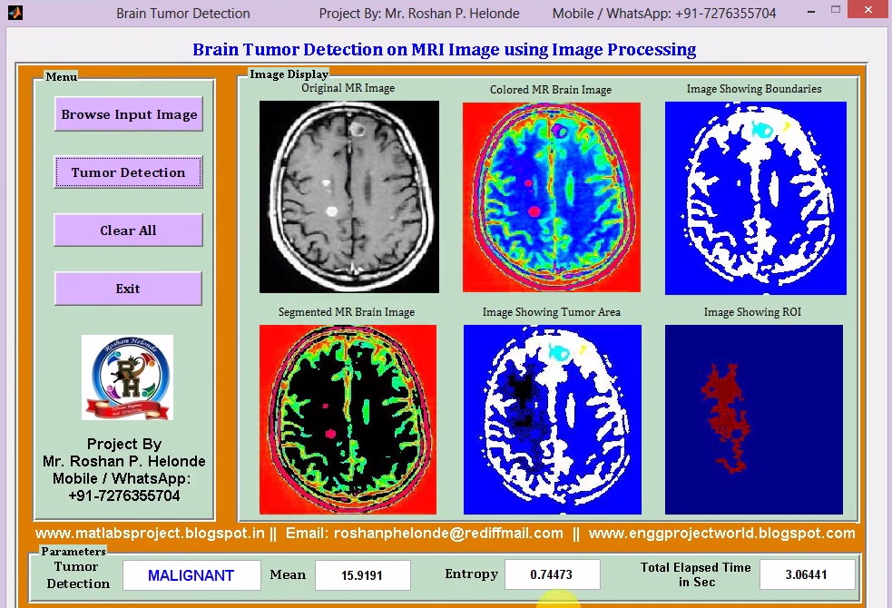
Brain Tumour Extraction from MRI Images Using MATLAB
The Brain Dynamics Toolbox provides an interactive simulation platform for exploring such systems in Matlab. It supports the major classes of differential equations that arise in computational neuroscience: Ordinary Differential Equations, Delay Differential Equations and Stochastic Differential Equations. The design of the graphical interface.

[PDF] Brain Tumour Extraction from MRI Images Using MATLAB Semantic Scholar
1 Link Muhammad, I use a program known as AFNI for such skull stripping. AFNI has been developed by the National Institute of Health (NIH) AFNI can be found here for download (it's free- http://afni.nimh.nih.gov/afni) and the part used for skull stripping is called 3dSkullStrip http://afni.nimh.nih.gov/pub/dist/doc/program_help/3dSkullStrip.html
Section 4.2 Example Box
We benchmarked four state-of-the-art rodent brain extraction methods, Rodent Brain Extraction Tool (RBET) (Wood et al. 2013), three dimensional pulse coupled neural networks. MSER was implemented by calling the MATLAB interface of the VLFeat package (version: 0.9.20).

Brain segmentation obtained with the Brain Extraction Tool (BET) from... Download Scientific
betsurf betsurf is the program that produces the three additional surfaces (inner & outer skull, outer scalp). It can output these surfaces as filled-in binary mask images, surface-only binary mask images and Geomview mesh format files. betsurf requires ideally good resolution T1- and T2-weighted input images.

I want to extract skull from Brain MRI usinG MORPHOLOGICAL OPERATIONS . But i didnt get a useful
The contour evolution tool is implemented in Matlab (MATLAB, 2010) and is also publicly available to researchers. Therefore, despite the recent progress in CT brain extraction, it is still challenging for a prospective researcher to distinguish the differences in performance of these tools and gauge the generalisability of these existing.

Brain Tumor Extraction From Mri Images Using Matlab Images Poster
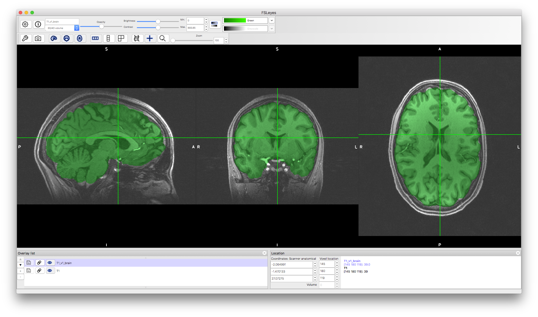
Chapter 1: Brain Extraction (also known as "skullstripping") Since fMRI studies focus on brain tissue, our first step is to remove the skull and non-brain areas from the image. FSL has a tool for this called bet, or the Brain Extraction Tool. It is the first button listed on the FSL GUI (indicated by "A" in the figure below).

Brain Tumor Extraction From Mri Images Using Matlab Images Poster
SPAMRI contains several features as follows: (1) open-source MATLAB-based package with a graphical user interface (GUI); (2) a set of images that can be generated for quality checking, such as Talairach transform, skull strip, and surface reconstruction; (3) user-friendly GUI with capabilities on statistical analysis, multiple comparison correct.
How to use the brain extraction tool
The contour evolution tool is implemented in Matlab (MATLAB, 2010) and is also publicly available to researchers. Therefore, despite the recent progress in CT brain extraction, it is still challenging for a prospective researcher to distinguish the differences in performance of these tools and gauge the generalisability of these existing.
(PDF) Brain Tumor Extraction from MRI Images Using MATLAB
In this tutorial we will discuss performing brain segmentation using the brain extraction tool (BET) in fsl and a robust version using a wrapper function in extrantsr, fslbet_robust. 1 Data Packages For this analysis, I will use one subject from the Kirby 21 data set.
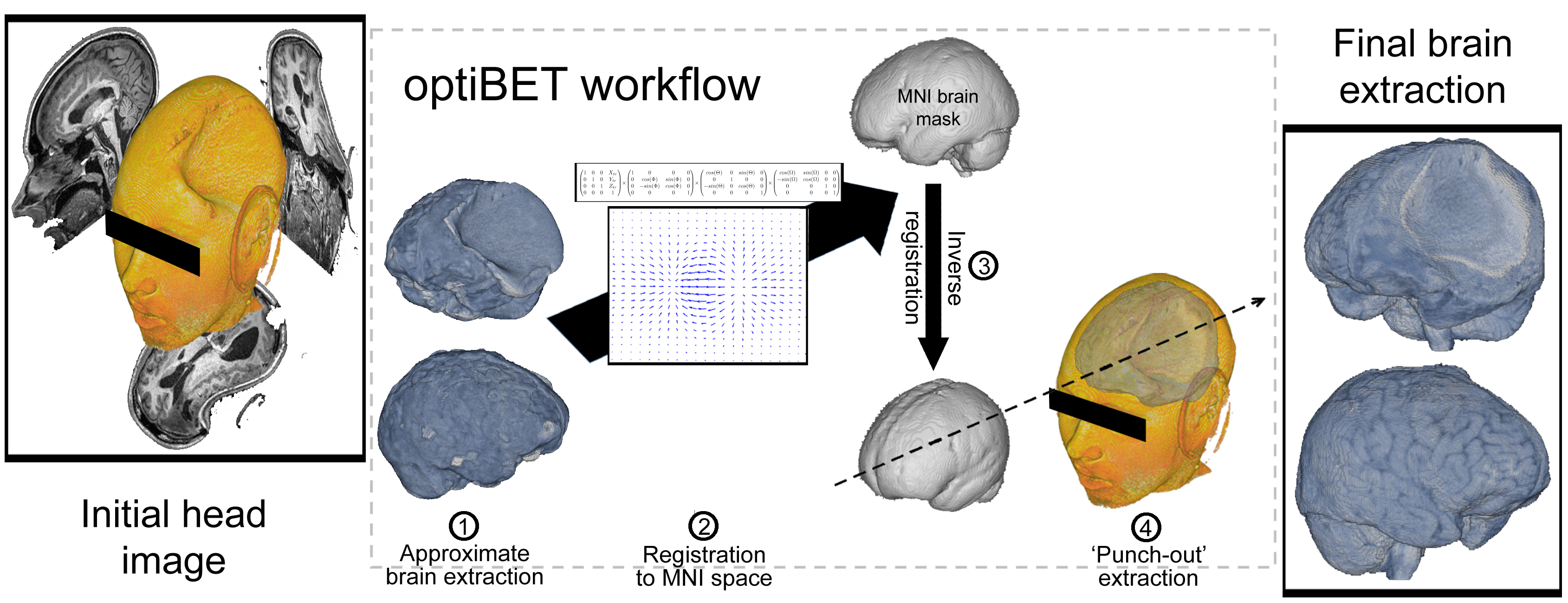
optiBET optimized brain extraction script for patient brain Professor Martin Monti's lab website
Brain extraction, also known as skull stripping, is a preliminary image post-processing technique that is fundamental for multiple applications in neuroscience and quantitative image analysis.

Brain Tumor Extraction From Mri Images Using Matlab Images Poster
The problem is that I am still a beginner with MATLAB, and the automatic segmentation does not allow me to have only the supratentorial part. So I don't know if there are other functions that allow me to segment the brain and have only the supratentorial part, to use it later as a mask to calculate the perfusion.